The Focus Microscope Camera
By Ken Roberts
Posted on 2014-04-03
While microscopes may open up dazzling possibilities for teachers to provide new understandings of the physical world, many science students often view microscopes with less enthusiasm. These youngsters have to set up slides, huddle around a limited number of microscopes, and take turns trying to see what was intended, fearing they may mistake air bubbles for single-cell organisms. Science lessons should foster curiosity and discussion, not worry.
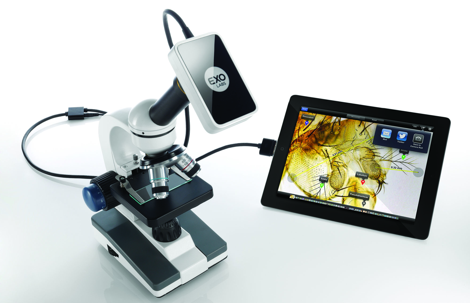
The new Focus Microscope Camera addresses the limitations of microscopes by connecting to the iPad, making microscopy more social, collaborative, and hands-on. The product connects virtually any microscope to the iPad, keeping both the camera and the tablet charged while in use. The kit includes the camera, microscope lens, AC adaptor, lens adapters, calibration card (for precise measurement), and 30-pin connector (the latest iPads and iPod Touches will need a lightning adaptor as well).
With the Focus Microscope Camera, magnified images are viewable in real time by students gathered around a shared iPad or by the whole class when the iPad view is projected wirelessly to a large screen. The Focus app (available for free download) allows students to collectively manipulate and examine the microscopic material. Additionally, the measurement and annotation tools enable them to document their findings. Included lens adapters make the camera compatible with any compound or dissecting microscope.
The product is easy to use for both teachers and students. The app offers a live interactive viewport, allowing still photos, video, time-lapse capture, fingertip point-to-point measurements, and pinch-finger zoom. And using the capabilities of the iPad, students can efficiently capture their observations, analyze their data, and share their results within a single class period.
The Focus app has precise tools. For example, students can easily examine hundreds of cells to determine the mitotic index of a tissue (e.g., onion root tips) and label the cells as mitotic or non-mitotic or by specific mitotic phases. In addition, students can study C. elegans (soil nematodes used for studying human disease) by accurately measuring the lengths of the worms to determine their life stage. Measurements can be done in units of microns, millimeters, or centimeters.
With the Focus Camera, teachers of all experience levels and disciplines can make lessons accessible and exciting while igniting student curiosity and encouraging active participation. The camera system was developed by two engineers in the medical instruments industry who recognized its potential in science education. The creators may not have realized how enabling the product would be for students with physical and visual disabilities.
Various accessories have been developed for the Focus Camera. An optional quick-change variable-focus lens turns the camera into a stand-alone benchtop science camera, useful for dissections, geology, ecology, forensics, and more. You will find this arrangement very useful once you realize how much your students appreciate seeing key images on a large monitor or TV.
The Focus Microscope Camera is an affordable, rugged, revolutionary technology that brings microscopy out of the dark ages from an isolated, often frustrating experience, to a 21st-century experience with active engagement, sharing, and collaboration in classrooms for students of all ages. This makes the Focus Microscope Camera an attractive and must-have technology in any science classroom setting.
Disclaimer: The views expressed in this blog post are those of the author(s) and do not necessarily reflect the official position of the National Science Teaching Association (NSTA).