Abstract
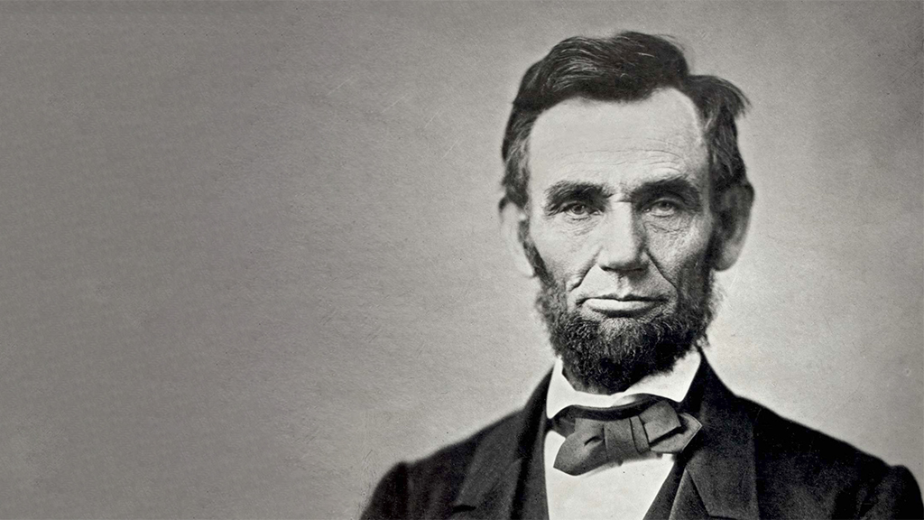
This case study, presented in the form of two mini-cases, is based on the story of Abraham Lincoln’s assassination by John Wilkes Booth. The first mini-case focuses on Lincoln. After learning about the medical details of the shooting, Lincoln’s injury, and the vigil kept by physicians until his death, students are asked to predict the brain structures damaged by the bullet and explain how increased intracranial pressure could produce many of the signs the physicians observed. The second mini-case focuses on Booth, who suffered a spinal cord injury when he was fatally wounded in the neck during his capture. Students use knowledge of spinal cord and cervical anatomy to connect Booth’s observed neurological deficits to autopsy records describing the tissue damage. Through both cases, students consider the functional anatomy of major brain and spinal cord structures. Originally written for an upper-level undergraduate neuroscience course for majors, the case could potentially be adapted for a non-neuroscience course, though if used in that context, it may require significant revisions depending on the material presented in that course.


